44 picture of respiratory system with labels
study the picture of the respiratory system below. Label the parts ... Science Elementary School answered study the picture of the respiratory system below. Label the parts correctly do this on your answer sheet Answer 0 vonembercachoremorin Answer: Number 1 nostrils number 2 mouth Advertisement Science A-Z Science Diagrams - Visual Teaching Tools Science Diagrams from Science A-Z provide colorful, full-page models of important, sometimes complex science concepts. Science Diagrams, available in both printable and projectable formats, serve as instructional tools that help students read and interpret visual devices, an important skill in STEM fields.
How the Lungs Work - The Respiratory System | NHLBI, NIH The main image shows the location of the lungs, pleura, and diaphragm. An inset image shows a closer view of the two layers of the pleura and the pleural space. The lungs are surrounded by the pleura, a membrane with two layers. The space between these two layers is called the pleural cavity.

Picture of respiratory system with labels
› current › title-42eCFR :: 42 CFR Part 84 -- Approval of Respiratory Protective ... (a) This section establishes a system under which NIOSH charges a fee for services provided to applicants for conformity assessment activities conducted by NIOSH for respiratory protective devices under 42 CFR part 84. This section specifies the purposes for which fees will be assessed and the cost factors for such assessments. Learning Task 2: Study the picture of the respiratory system below ... Learning Task 2: Study the picture of the respiratory system below. Label the parts correctly. - 24101441 Female Reproductive System - Anatomy Pictures and Information - Innerbody The female reproductive system includes the ovaries, fallopian tubes, uterus, vagina, vulva, mammary glands and breasts. These organs are involved in the production and transportation of gametes and the production of sex hormones. The female reproductive system also facilitates the fertilization of ova by sperm and supports the development of ...
Picture of respiratory system with labels. pressbooks.uwf.edu › chapter › cardiovascular-systemCardiovascular System – Medical Terminology for Healthcare ... We can detect and record the electrical activity of the heart’s conduction system using an electrocardiogram (ECG or EKG). Figure 9.7 shows the electrical impulse originating in the SA node (step 2) and traveling through the heart’s conduction system, allowing the heart to complete one cardiac cycle. Each waveform on the ECG tracing ... Equine Veterinary Products & Equipment | Patterson Vet Take advantage of exclusive 0% summer financing rates from Patterson Veterinary and get the equipment you’ve had your eye on. Add services to your practice with new ultrasound equipment. Your patients will be the picture of health. Offer ends August 31, 2022. View details Home: The Histology Guide The site is divided into topics, which may be worked through in any order. You can see histological slides on the pages and can turn labels on or off to help them identify features. In some cases, there is a section like a 'virtual microscope' - you can scan around a large picture using the mouse and try to identify features. Digestive System Label Diagram - digestive system coloring page az ... Digestive System Label Diagram - 16 images - digestive system key terms flashcards quizlet, digestive system diagram with labels and functions news word, anatomy digestive system assignments mr lyons science, digestive system for kids how digestion works human body parts for,
E Learning Task 2: Study the picture of the respiratory system below ... The respiratory system is one of the main organ systems of the human body. The most important organ in this system is the lungs, which helps us breathe. The lungs disperse the oxygen that we take from the air outside into the hearts and the blood vessels, which will go to the brain. Without the respiratory system, we will not be able to breathe. Respiratory System Quiz: Questions With Answers - ProProfs Small air sacs in the lungs where many capillaries exchange carbon dioxide for oxygen are: 4. 5. Small spaces in the skull thought to regulate the temperature and humidity of the air taken into the body. 6. 7. This tube carries air down to the windpipe. 8. Anatomy Chart - How to Make Medical Drawings and Illustrations Anatomy Chart What is an Anatomy Chart? An anatomy chart refers to a visual depiction of the human body. It can show the entire body or focus on a particular system using systemic anatomy such as the muscular, skeletal, circulatory, digestive, endocrine, nervous, respiratory, urinary, reproductive, and other systems. There are many different branches of anatomy … Amazon.com: ChildLife Essentials Organic Vitamin D3 - Vitamin D … Buy ChildLife Essentials Organic Vitamin D3 - Vitamin D Drops for Kids, Supports Immune, Respiratory, Heart, & Bone Health, All-Natural, Gluten-Free, Non-GMO - Natural Berry Flavor, 1 Fl Oz Bottle (Pack of 2) on Amazon.com FREE SHIPPING on qualified orders
› content › 1Respiratory Illnesses: 13 Types of Lung Infections - OnHealth Apr 08, 2022 · Upper Respiratory Infection. Types of upper respiratory infection include the common cold (head cold), the mild flu, tonsillitis, laryngitis, and sinus infection. Of the upper respiratory infection symptoms, the most common is a cough. Lung infections may also lead to a stuffy or runny nose, sore throat, sneezing, achy muscles, and headache. Male Reproductive System - Explore Anatomy with Detailed Pictures Male Reproductive System. The male reproductive system includes the scrotum, testes, spermatic ducts, sex glands, and penis. These organs work together to produce sperm, the male gamete, and the other components of semen. These reproductive organs also work together to deliver semen out of the body and into the vagina where it can fertilize egg ... 9 Cardiovascular System - University of West Florida Respiratory System. 13. Digestive System. 14. Endocrine System. 15. Urinary System. 16. Male Reproductive System. ... A computer picture of the heart created by bouncing high-energy sound waves ... The sagittal view labels read (from top, clockwise): first rib, aortic arch, thoracic arch, esophagus, inferior vena cava, diaphragm, thymus, trachea. › main › resourcetypeScience A-Z Science Diagrams - Visual Teaching Tools Science Diagrams from Science A-Z provide colorful, full-page models of important, sometimes complex science concepts. Science Diagrams, available in both printable and projectable formats, serve as instructional tools that help students read and interpret visual devices, an important skill in STEM fields.
Respiratory System
Mild respiratory COVID can cause multi-lineage neural cell and … 13.06.2022 · Mild respiratory COVID causes neuroinflammation and multi-lineage cellular dysregulation in the central nervous system, a phenomenon mirroring cancer-therapy-related cognitive impairment.

Respiratory System Labeled Vector de stock (libre de regalías)119610076; Shutterstock
Free Respiratory System Worksheets and Printables - Homeschool Giveaways Respiratory System Doodle Labeled Coloring Page - This coloring page includes wonderful details about the respiratory system such as an explanation about how the diaphragm contracts and a close-up image of the lung alveoli. If your kids love to color, this is the perfect worksheet for you! Respiratory System Notebooking Pages

lpcomputerlab: Grade 4- Respiratory System
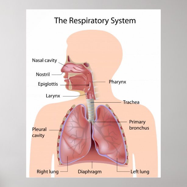
Respiratory System Organs and Their Functions - New Health Advisor The airways (nose, mouth, pharynx, larynx etc.) allow air to enter the body and into the lungs. The lungs work to pass oxygen into the body, whilst removing carbon dioxide from the body. The muscles of respiration, such as the diaphragm, work in unison to pump air into and out of the lungs whilst breathing. Physiology of Gas Exchange
Respiratory System | ClipArt ETC
Histology, Respiratory Epithelium - StatPearls - NCBI Bookshelf The respiratory system is constantly filtering through the external environment as humans breathe air. The airways must maintain the ability to clear inhaled pathogens, allergens, and debris to maintain homeostasis and prevent inflammation. The respiratory system subdivides into a conducting portion and a respiratory portion.

Circulation and respiration | Circulatory and respiratory systems | Siyavula
Respiratory system quizzes and labeled diagrams | Kenhub Take a look at the labeled diagram of the respiratory system above. As you can see, there are several structures to learn. Spend a few minutes reviewing the name and location of each one, then try testing your knowledge by filling in your own diagram of the respiratory system (unlabeled) using the PDF download below. Respiratory system unlabeled

Human Anatomy Lab: The Urinary and Reproductive Systems
30 Fun And Interesting Respiratory System Facts For Kids - MomJunction The Respiratory Tract Image: Shutterstock The respiratory tract is divided into two sections, namely, upper and lower. The part above the voice box or larynx is upper respiratory tract and the one below it is lower respiratory tract. The respiratory tract is lined by respiratory mucosa or respiratory epithelium (2).
Respiratory System Posters, Prints & Poster Printing | Zazzle CA
Respiratory system: Anatomy and functions | Kenhub The tracheobronchial tree is a portion of the respiratory tract that conducts the air from the upper airways to the lung parenchyma. It consists of the trachea and the intrapulmonary airways (bronchi and bronchioles).The trachea is located in the superior mediastinum and represents the trunk of the tracheobronchial tree.
Post a Comment for "44 picture of respiratory system with labels"