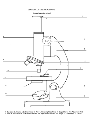
40 microscope diagram without labels
7th grade Science - Microscope Diagram - Quizlet The Parts of a Microscope. 12 terms. totobear PLUS. Sets found in the same folder. Science Key terms 7th grade. 13 terms. palocastillo. 7th Grade Earth Science. 9 terms. EliseC17. 7thGrade Review - Cells/Biology. 26 terms. SolizScience TEACHER. 7th grade Science, Cell theory. 8 terms. Super1412. Other sets by this creator. PDF Label compound microscope worksheet - Weebly [clearBoth] [clearBoth] Microscope diagram without label After you've studied all the pieces of the composite microscope, it's time to put your brain to the test. Print an unmarked microscope chart and check that you can fill out all the blanks. [clearBoth] [clearBoth] Blank microscope diagram Next we have an empty microscope diagram.
Compound Microscope Parts - Labeled Diagram and their ... The eyepiece (or ocular lens) is the lens part at the top of a microscope that the viewer looks through. The standard eyepiece has a magnification of 10x. You may exchange with an optional eyepiece ranging from 5x - 30x. [In this figure] The structure inside an eyepiece. The current design of the eyepiece is no longer a single convex lens.

Microscope diagram without labels
Diagram of a Compound Microscope - Biology Discussion Information recorded on adhesive label is stuck to the base of the microscope for future reference. (ii) Use: Having calibrated the eyepiece scale for all the objective lenses on the microscope, one can use it to measure the dimensions of cellular and sub-cellular structures, e.g., bacterial cells, fungal spores onion epidermal cells etc. Label the microscope - Science Learning Hub Jun 8, 2018 — Use this interactive to identify and label the main parts of a microscope. Drag and drop the text labels onto the microscope diagram.Labels: DescriptionEye piece lens: The lens you look through – no...Light source: Sends light onto the specimen/slideCoarse focus adjustment: Moves the lens up or ... Microscope, Microscope Parts, Labeled Diagram, and Functions The Iris Diaphragm is located above the condenser lens and below the microscope stage. The different sized holes in the diaphragm helps to vary the size of the cone and intensity of light that is projected upward into the slide. However, there is no set rule regarding which setting to use for a particular power.
Microscope diagram without labels. A Study of the Microscope and its Functions With a Labeled ... Here, unlabeled microscope diagrams have been provided for your perusal, which will help you practice and test your understanding of the instrument. Types of Microscopes Depending on the source of illumination, microscopes can be divided into two categories. They are: PDF Parts of the Light Microscope - Science Spot B. NOSEPIECE microscope when carried Holds the HIGH- and LOW- power objective LENSES; can be rotated to change MAGNIFICATION. Power = 10 x 4 = 40 Power = 10 x 10 = 100 Power = 10 x 40 = 400 What happens as the power of magnification increases? Microscope Labeling - The Biology Corner Students label the parts of the microscope in this photo of a basic laboratory light microscope. Can be used for practice or as a quiz. Name_____ Microscope Labeling . Microscope Use: 15. When focusing a specimen, you should always start with the _____ objective. Labeling Microscope Worksheet | Teaching Resources A straightforward worksheet in which students are required to identify the parts of a basic microscope. Tes classic free licence. Reviews. 4.7 Something went wrong, please try again later. MACS0647-JD. a year ago. report. 5. Thanks. Very helpful. Empty reply does not make any sense for the end user ...
Amazing 27 Things Under The Microscope With Diagrams Amazing 27 Things Under The Microscope With Diagrams February 20, 2022 by Anupama Sapkota Note: Each image source is given below in this post of respective subheadings. Table of Contents 1. Amoeba under the microscope Direct observation Observation after staining 2. Algae under the microscope Chlorophyta Chromophyta Cryptophyta Rhodophyta Microscope With Labels Clip Art at Clker.com - vector clip ... diagram of a microscope; sketch of a compound microscope; diagram of microscope with labels; sketch of compound microscope; picture of microscope without label; a microscope without labels; compound microscope outline; microscope drawing no labels; microscope side view; draw and label the parts of a microscope; microscope no labels; parts of ... Compound Microscope Parts, Functions, and Labeled Diagram Compound Microscope Definitions for Labels. Eyepiece (ocular lens) with or without Pointer: The part that is looked through at the top of the compound microscope. Eyepieces typically have a magnification between 5x & 30x. Monocular or Binocular Head: Structural support that holds & connects the eyepieces to the objective lenses. 16 Parts of a Compound Microscope: Diagrams and Video Once you have an understanding of the parts of the microscope it will be much easier to navigate around and begin observing your specimen, which is the fun part! The 16 core parts of a compound microscope are: Head (Body) Arm Base Eyepiece Eyepiece tube Objective lenses Revolving Nosepiece (Turret) Rack stop Coarse adjustment knobs
PDF Label parts of the Microscope: Answers Label parts of the Microscope: Answers Coarse Focus Fine Focus Eyepiece Arm Rack Stop Stage Clip . Created Date: 20150715115425Z ... PDF Parts of a Microscope Printables - Homeschool Creations Label the parts of the microscope. You can use the word bank below to fill in the blanks or cut and paste the words at the bottom. ... without needing to move the microscope ? the head •What is the magnification level on the eyepiece of a microscope?10x (see objective Label the Microscope Diagram | Download Scientific Diagram Download scientific diagram | Label the Microscope Diagram from publication: Laboratory Exercises in Microbiology: Discovering the Unseen World through Hands-on Investigation | Microbiology ... Label Microscope Diagram - EnchantedLearning.com Label Microscope Diagram Using the terms listed below, label the microscope diagram. Inventions and Inventors: arm - this attaches the eyepiece and body tube to the base. base - this supports the microscope. body tube - the tube that supports the eyepiece. coarse focus adjustment - a knob that makes large adjustments to the focus.
Natural Sciences Grade 9
Labelled Diagram of Compound Microscope - Biology Discussion The below mentioned article provides a labelled diagram of compound microscope. Part # 1. The Stand: The stand is made up of a heavy foot which carries a curved inclinable limb or arm bearing the body tube. The foot is generally horse shoe-shaped structure (Fig. 2) which rests on table top or any other surface on which the microscope in kept.

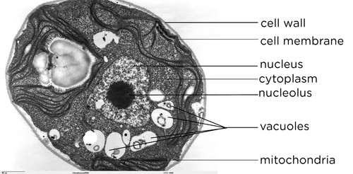
A typical animal cell (as seen in an electron microscope) Medical Ima…
Microscope Label Interactive Worksheets & Teaching ... Microscope Interactive Notebook Activity by Jodi's Jewels 12 $1.89 PDF Students will complete a timeline of the history of the microscope, label a diagram, and create a pocket foldable with terms and definition cards. The timeline can be completed according to the teacher's directions or like the answer key example.

Labelled Microscope Diagram Gcse - Micropedia
Labeling the Parts of the Microscope | Microscope World ... Labeling the Parts of the Microscope This activity has been designed for use in homes and schools. Each microscope layout (both blank and the version with answers) are available as PDF downloads. You can view a more in-depth review of each part of the microscope here. Download the Label the Parts of the Microscope PDF printable version here.

Simple Microscope Labeled Diagram - Micropedia
Simple Microscope - Parts, Functions, Diagram and Labelling A simple microscope is a device that only has one lens for magnification. It functions the same way as the magnifying glass. Although it is simple in terms of design and function, it is useful I various fields including medicine, jewelry and watchmaking, and agriculture, to name a few. References
Label a microscope - Teaching resources
Microscope Labeling Game - PurposeGames.com About this Quiz. This is an online quiz called Microscope Labeling Game. There is a printable worksheet available for download here so you can take the quiz with pen and paper. This quiz has tags. Click on the tags below to find other quizzes on the same subject. Science.

Rens blog : Science, cells
Microscope Parts and Functions With Labeled Diagram and ... First, the purpose of a microscope is to magnify a small object or to magnify the fine details of a larger object in order to examine minute specimens that cannot be seen by the naked eye. Here are the important compound microscope parts... Eyepiece: The lens the viewer looks through to see the specimen.

A schematic diagram of the light path in the ptychographic microscope... | Download Scientific ...
Compound Microscope: Definition, Diagram, Parts, Uses ... The compound microscope is mainly used for studying the structural details of cell, tissue, or sections of organs. The parts of a compound microscope can be classified into two: Non-optical parts Optical parts Non-optical parts Base The base is also known as the foot which is either U or horseshoe-shaped.
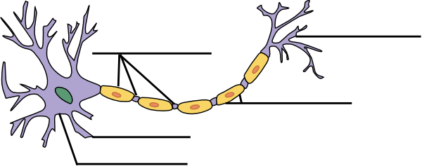
Label The Neuron Clip Art at Clker.com - vector clip art online, royalty free & public domain
Animal Cell Simple Labeled Diagram - Mitosis Diagram ... Cells communicate with one another and are responsible for transmitting microscope label the diagram of a microscope. As observed in the labeled animal cell diagram, the cell membrane forms the confining factor of the cell, that is it envelopes the cell constituents together and gives the cell its shape, form, and existence.
Microscope Diagram - Fill Online, Printable, Fillable, Blank | PDFfiller
Parts of a microscope with functions and labeled diagram Figure: Diagram of parts of a microscope There are three structural parts of the microscope i.e. head, base, and arm. Head - This is also known as the body. It carries the optical parts in the upper part of the microscope. Base - It acts as microscopes support. It also carries microscopic illuminators.

Biology: Soalan SPM Chapter 2
Microscope Lab Microscope Diagram. History of the microscope. Microscope Information. Questions: 1. Did the letter appear in the same orientation when viewed through the microscope as viewed without the microscope? 2. When you move the slide to the right what direction does it appear to move under the microscope? 3. What happened to the image when you ...
Unlabeled Microscope Diagram - Cliparts.co
Microscope, Microscope Parts, Labeled Diagram, and Functions The Iris Diaphragm is located above the condenser lens and below the microscope stage. The different sized holes in the diaphragm helps to vary the size of the cone and intensity of light that is projected upward into the slide. However, there is no set rule regarding which setting to use for a particular power.

with a neat labelled diagram explain the formation of image in a compound microscope - Brainly.in
Label the microscope - Science Learning Hub Jun 8, 2018 — Use this interactive to identify and label the main parts of a microscope. Drag and drop the text labels onto the microscope diagram.Labels: DescriptionEye piece lens: The lens you look through – no...Light source: Sends light onto the specimen/slideCoarse focus adjustment: Moves the lens up or ...
(22).jpg)
An Ultimate Quiz On Microscope Parts And Functions! - ProProfs Quiz
Diagram of a Compound Microscope - Biology Discussion Information recorded on adhesive label is stuck to the base of the microscope for future reference. (ii) Use: Having calibrated the eyepiece scale for all the objective lenses on the microscope, one can use it to measure the dimensions of cellular and sub-cellular structures, e.g., bacterial cells, fungal spores onion epidermal cells etc.
Labelling a Microscope - Labelled diagram
Microscope Labeling Game Answers | Games World
Labeled-microscope-diagram

Onion Epidermal Cell Labeled Diagram - Diagram For You
Post a Comment for "40 microscope diagram without labels"